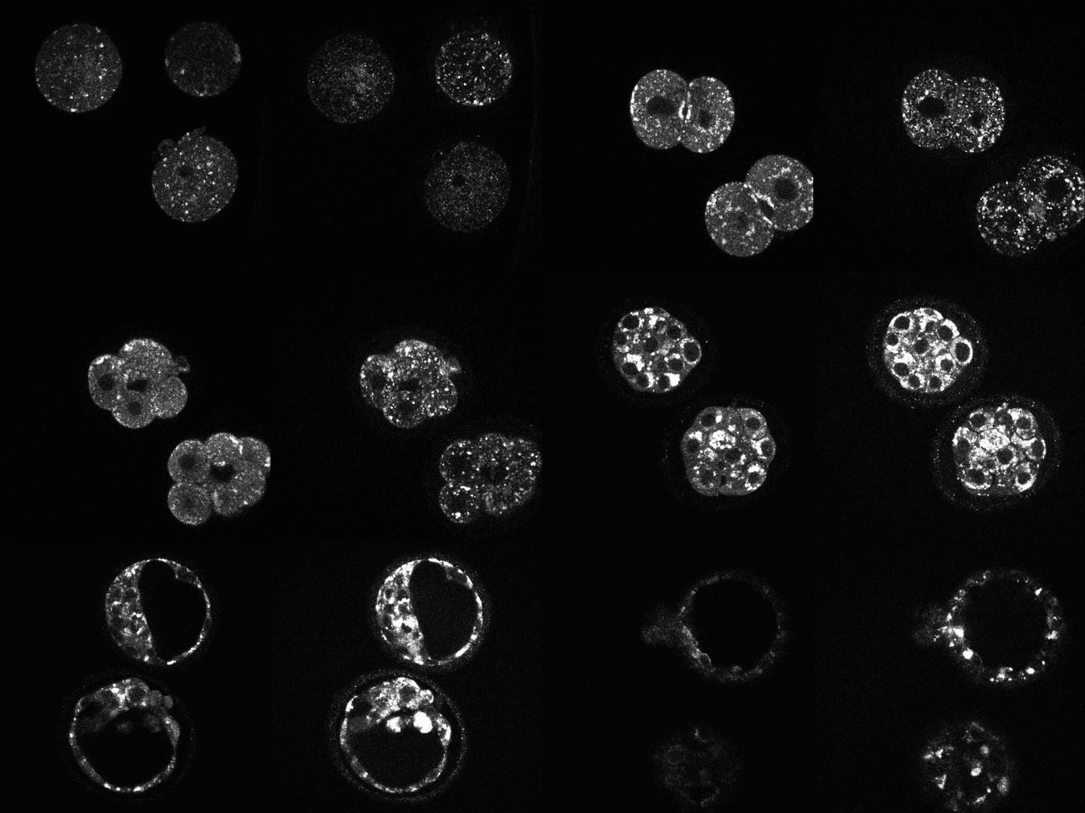
Early human embryos are self-organizing systems. After fertilization, the resulting embryo undergoes a series of cell divisions. After approximately four days of this “cleavage stage,” the embryo compacts, as the cells firmly adhere to each other, at which time it is referred to as a “morula.” A fluid cavity then forms inside the embryo, at which point it is called a “blastocyst.” Cells in the blastocyst differentiate into either the inner cell mass (which will develop into the fetus) or the trophectoderm (which will go on to become the embryonic part of the placenta). The embryo then implants into the endometrium of the uterus. Errors in chromosome segregation are the primary cause of age-related infertility in women. Preimplantation embryo development occurs with almost no change in mass. Thus, all these early developmental events largely consist of a reorganization of the same material that was present in the unfertilized egg.
We are studying cell division and metabolism in mouse and human preimplantation embryos, human oocytes, and human cumulus cells (which are physically and metabolically associated with oocytes). We are performing statistical analysis of massive data sets from in-vitro fertilization (IVF) clinics to learn more about the self-organization of preimplantation human embryos. We are investigating the hypothesis that chromosome segregation errors in oocytes and early embryos may be caused by metabolic defects. We are also attempting to use quantitative approaches to improve clinical IVF treatments of human infertility.